3-D Ultrasound Quantifies Plaque Burden in Healthy Middle-Aged Cohort
Even at midlife, 55% of men and 30% of women have subclinical atherosclerosis identified on ultrasound, report investigators.

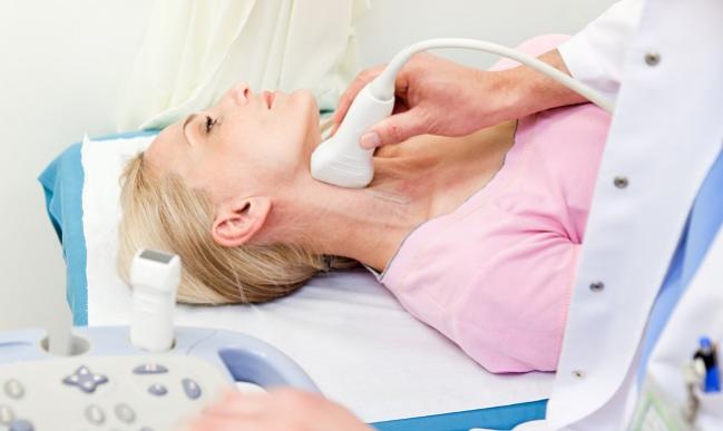
The use of 3-D vascular ultrasound of the carotid and femoral arteries can safely quantify early atherosclerotic disease burden in healthy men and women, according to the results of a new study.
Imaging with 3-D ultrasound identified a higher burden of atherosclerotic plaque in men compared with women and an increasing plaque burden as individuals aged. Additionally, plaque burden, specifically in the femoral artery, strongly correlated with cardiovascular disease risk factors.
The new study, which was led by Beatriz López-Melgar, MD (Centro Nacional de Investigaciones Cardiovasculares Carlos III, Madrid, Spain) and Valentin Fuster, MD (Icahn School of Medicine at Mount Sinai, New York, NY), and published July 10, 2017, in the Journal of the American College of Cardiology, provides “evidence that 3-D vascular ultrasound is a feasible and reproducible approach to not only detect, but also quantify, early atherosclerotic burden,” according to the researchers.
Plaque volume, they explain, is a comprehensive measurement that includes disease presence and amount, which likely better reflects the cumulative effects of chronic cardiovascular risk factor exposure. The group points out that while 2-D vascular ultrasound can detect and grade the extent of atherosclerosis in different territories, 3-D ultrasound has been proposed as a better means to quantify peripheral atherosclerosis.
The PESA Study
In this population-based observational study, the researchers evaluated the plaque burden in 3,820 healthy men and women aged 40 to 54 years participating in the Progression of Early Subclinical Atherosclerosis (PESA) study. In addition to a physical examination, lifestyle questionnaires, and bloodwork, individuals had a baseline 3-D ultrasound of the bilateral femoral and carotid arteries. Follow-up visits are scheduled at 3 and 6 years.
At the baseline visit, the median global plaque burden was 50.8 mm3 and was higher in men than in women (63.4 mm3 vs 25.7 mm3; P < 0.001). Overall, atherosclerosis was present in 55.8% of men and 30.5% of women, was more pronounced in the femoral artery than in the carotid, and increased significantly with age.
Atherosclerotic plaque burden also tracked with cardiovascular risk factors. In a multivariate-adjusted analysis, male sex, increasing age, smoking (10 pack-years), dyslipidemia, and hypertension were associated with plaque burden. Diabetes was associated with plaque burden in the femoral but not carotid artery.
Gary Mintz, MD (Cardiovascular Research Foundation, New York, NY), who was not involved in the study, told TCTMD the imaging findings as a whole were not too surprising, particularly since it’s been long documented that Western populations have a high prevalence of atherosclerosis.
“This study extends that to midlife,” said Mintz. “If you look at plaque burden, global plaque burden of 30%, 40%, 50% is not that unusual and it increases as people get older. The Framingham study showed that a long time ago—older people have more coronary disease, and men have more than women until women get older. This is perhaps a more novel way of looking, at but it’s not too surprising.”
Regarding the technology, Mintz said one of the limitations of 2-D vascular ultrasound is the inability to sample a significant proportion of the vasculature. Also, one of the problems with ultrasound, in general, remains the reproducibility of image acquisition, he said.
In the present study, reproducibility was assessed as the agreement between three readers for global plaque burden, but Mintz said he would have liked to see a reproducibility analysis based on the technical aspects of image acquisition. External ultrasound is more dependent, or at least has the potential to be more dependent, on these technical aspects, including the skills of the technologist, he said.
“Since ultrasound is 100% noninvasive, it would have been better to have the reproducibility analysis started with acquiring a second set of images,” said Mintz. “For example, technician 1 leaves the room, technician 2 comes in and redoes the study. Now, that’s kind of arcane but it’s important because anytime you’re doing serial studies, or studies amongst many patients, then reproducibility is really important.”
Potential Clinical Implications
Regarding the clinical implications, Mintz said there is reason to think 3-D ultrasound will supplement 2-D ultrasound but it depends a lot on the cost of the equipment as well as technical training. In terms of how it may be used, he said, it’s a matter of philosophy.
“If you are of the belief that atherosclerotic coronary disease is ubiquitous in the Western population and everybody should be taking a statin, then it’s a little unclear what this would mean clinically,” he said. That said, 3-D ultrasound could be used to screen for individuals who have aggressive, albeit asymptomatic, atherosclerosis in order to target them with more intensive therapy, such as a PCSK9 inhibitor.
In an editorial accompanying the study, J. David Spence, MD (Western University, London, Canada), points out that total plaque burden is a much stronger predictor of cardiovascular risk than ultrasound measurements of intima-media thickness (IMT) and is more sensitive to the effects of therapy.
For example, in the High-Risk Plaque Study, carotid plaque burden was correlated with coronary calcium scores and predictive of adverse events, but IMT was not, he notes. Spence states that the development of 3-D ultrasound will make it easier to implement imaging studies where the artery is treated rather than simply treating cardiovascular disease risk factors.

Michael O’Riordan is the Managing Editor for TCTMD. He completed his undergraduate degrees at Queen’s University in Kingston, ON, and…
Read Full BioSources
López-Melgar B, Femández-Friera L, Oliva B, et al. Subclinical atherosclerosis burden by 3-D ultrasound in mid-life. J Am Coll Cardiol. 2017;70:301-313.
Spence JD. Approaching automated 3-dimensional measurement of atherosclerotic plaque volume. J Am Coll Cardiol. 2017;70:314-317.
Disclosures
- The study authors report no conflicts of interest.
- Mintz is the chief medical officer of the Cardiovascular Research Foundation, the publisher TCTMD.
Comments