Atherosclerosis Seen in 500-Year-Old Inuit Mummies
CT scanning revealed specks of calcium in three of these people, who would have eaten a diet rich in omega-3 fatty acids.

Building on prior research on vascular calcification in the mummified remains of ancient people, the Horus Study Group has now made a similar discovery in Inuit people who lived on the Greenlandic island of Uunartoq in the 1500s.
In three of four mummies preserved primarily by the cold environment, whole-body CT scanning revealed specks of calcification in the arterial tree, including in the carotids and the distal aorta.
“Incomplete visualization of the arterial vascular tree precluded accurate grading of the magnitude or severity of vascular [calcification] and evaluation of clinical disease,” researchers led by L. Samuel Wann, MD (Ascension Healthcare, Milwaukee, WI), report. “Nevertheless, the appearance of vascular calcification in these three mummies resembled previous observations of atherosclerosis in mummies and living humans.”
Speaking with TCTMD, Wann noted that the Inuit people were hunter-gatherers with an active lifestyle and a marine-based diet rich in omega-3 fatty acids. While it might be surprising to some that evidence of atherosclerosis was found in such a population, Wann said, “atherosclerosis is a multifactorial disease.”
Previously, the Horus researchers have shown that atherosclerosis existed even further back in history—at least as long ago as 4,000 BCE—among people from various civilizations, including the Egyptians, Peruvians, Ancestral Puebloans, and Unangan hunter-gatherers. But none of those populations ate a primarily marine-based diet, which might be expected to protect against atherosclerosis.
For this new study, published recently as a research letter in JAMA Network Open, the investigators examined five Greenlandic Inuit mummies that were discovered in 1929 and were being kept at the Peabody Museum of Archaeology and Ethnology in Cambridge, MA. “Grave goods and typical clothing indicated burial in the 1500s, when these individuals would have lived in stone, whale bone, and seal skin huts and hunted from kayaks with spears, bows, and arrows for their diet of fish, birds, marine mammals, and caribou,” the authors note.
The mummies included one infant and, based on bone and dental development, four teens/young adults—two men ages 18 to 30 and two women ages 16 to 30 at the time of their deaths from unknown causes.
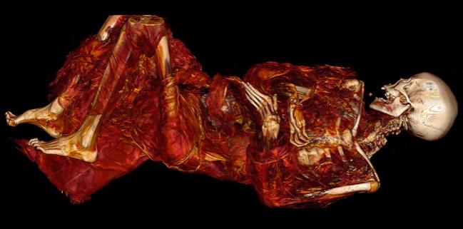
The remains were transported to Brigham and Women’s Hospital in Boston, MA, to undergo multidetector, whole-body CT scanning. The infant was excluded from the analysis due to a “paucity of nonbony tissue.” As for the other mummies, “remnants of the carotid arteries, the thoracic and retroperitoneal aorta, and iliac arteries were preserved in all four individuals, but reliable anatomic landmarks within the heart could not be identified,” Wann et al report. Calcification was seen in three cases.
Asked about whether the calcification definitively indicates atherosclerosis, Wann said that the research group’s experience with other mummies supports that it does. “We’re not absolutely certain we have vascular calcification, but we’re pretty certain,” he said.
What’s not known is whether these people had clinical heart disease. Prior work has shown that some Egyptian mummies with calcification did have signs of clinical disease, but Wann pointed out that “we’re talking about vascular calcification, which even in living humans . . . doesn’t mean you’ve had a heart attack or have occlusive disease. It means you have atherosclerosis and you have a calcified form of it.”
The message Wann takes away from this and other studies of vascular calcification in mummies is that atherosclerosis does not have just one cause. That these Inuit people, who had an active lifestyle and ate a diet high in fish oil, still had atherosclerosis begs the question: why? One potential explanation, according to Wann, is the burning of fires in igloos where the Inuit people lived. Exposure to particulate matter—from fires and other sources—has been recognized as a factor contributing to the development of atherosclerosis.
“Reducing particulate emissions by gasoline- and diesel-powered cars is a major initiative of people involved in preventive health,” Wann said.
That said, it’s unknown whether exposure to fires is what led to the calcification observed in these mummies, he added. “What we do know is that they had some vascular calcification and several hundred years ago in a preindustrial society atherosclerosis did exist.”
Photo Credit: Horus Research Team. CT scout image of a Greenlandic mummy prior to diagnostic processing.

Todd Neale is the Associate News Editor for TCTMD and a Senior Medical Journalist. He got his start in journalism at …
Read Full BioSources
Wann LS, Narula J, Blankstein R, et al. Atherosclerosis in 16th-century Greenlandic Inuit mummies. JAMA Network Open. 2019;2(12):e1918270.
Disclosures
- The Paleocardiology Foundation provided funds to transport the mummies from the Peabody Museum to Brigham and Women’s Hospital, where CT scanning was performed without charge.
- Wann reports no relevant conflicts of interest.
Comments