Blasting Off: Experience Grows for Intravascular Lithotripsy for Coronary Plaque
Future head-to-head studies are needed, experts say, noting that despite the excitement, the technology has certain disadvantages to consider.

WASHINGTON, DC—Early experience with intravascular lithotripsy (IVL) to modify severe coronary calcium before stent implantation shows that the technology is a safe and feasible alternative to currently available techniques, according to new data.
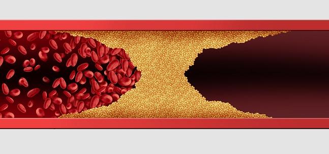
The lithotripsy system (Shockwave Medical; Fremont, CA) is made up of a semicompliant balloon catheter, connector cable, and generator. When deployed, it releases pulsatile mechanical energy that causes microfractures in the calcium. In 2017, the system showed promise in coronary lesions in the DISRUPT CAD, though it has not yet been compared with other plaque modification methods like rotational and orbital atherectomy.
“There are many advantages of IVL over other modalities, including being able to provide a more controlled means of calcium modification; it avoids the no reflow that we see in rotational atherectomy that can put patients at high risk; it allows continuous simultaneous guidewire position for bifurcation intervention, particularly left main stem; . . . and it has the ability to modify calcification without injury to the intimal soft tissue,” said Julian Yeoh, MBBS (King’s College Hospital, London, England), who presented findings from a new study last week at CRT 2019.
Yeoh and colleagues looked at 54 procedures using lithotripsy—14 study cases similar to those in DISRUPT CAD and 40 clinical cases—from their institution between May 2016 and September 2018. The indication for lithotripsy was based on the presence of an undilatable lesion, stent underexpansion, or severe calcification on either angiography or intravascular imaging. Almost half (48%) of the clinical patients presented with ACS, while 13% had renal replacement therapy and 28% had left main stem and multivessel intervention. Overall, 11% had a previous undilatable lesion.
Total procedural success was 91%, and stent delivery was possible in all cases. There were no instances of device-related coronary perforation, no reflow, ventricular arrythmia, or 30-day target lesion failure.
Among the 52% of cases with available optical coherence tomography imaging data, there was no statistically significant difference between the study and the clinical case arms regarding post-PCI main lumen area (4.2 vs 4.3 mm; P = 0.83) or main residual area stenosis (21.9% vs 19.0%; P = 0.55).
Yeoh said the main disadvantage of IVL “is that it can be quite a bulky balloon and may require GuideLiner or heavy wire to deliver the device to the target lesion.” However, the latest iteration, the C2 balloon, is a bit easier to deliver to the target lesion, he added.
Moving Forward With IVL
In discussion following his presentation, Yeoh noted that “at the moment, there's not many centers using the technology. Obviously, our center has vast experience.” Moving forward, “one of the things I think we need to be careful of is understanding the place of each modality. There is a place for rotational atherectomy, there is a place for orbital atherectomy, there's a place for the cutting balloon,” he said. “[IVL is] gathering momentum. I think we need more evidence to suggest that it’s safe in particular scenarios.”
Session co-moderator Carlos Sanchez, MD (OhioHealth-Riverside Methodist Hospital, Powell, OH), asked if there would be “any difference in using it in patients with concentric calcification and very eccentric calcification.”
“I think that IVL will actually be the modality of choice for concentric calcification,” Yeoh responded. “If you think about rotational atherectomy and how it works, if you have concentric calcification, all you're doing is boring a hole where the wire is biased. That may not deal with the lesion and prepare it adequately because you still might not be able to prep that calcium if it's biased to one side.”
Citing no known contraindications for IVL, Yeoh said its main advantage “is that it has a very short learning curve, so I think it can be used in, say, your county hospital where surgical backup may not be available. I think the applications are coming, and yes, we need more research, but . . . I think it's going to be something to watch.”
‘Simple, Easy to Use’
Showing similar enthusiasm for IVL, Richard Anderson, MD (Cardiff and Vale University Health Board, Wales), who spoke on the technology in a hot topics session also at CRT 2019, said, “I think intravascular imaging is crucial and almost mandatory for all of our severe calcific cases, and lithotripsy has a clear role here. Imaging does show us the limitations of our current devices. It shows us how superficially it really ablates calcium within the arteries and it really doesn't deal with the more concentric and deeper calcium. This is where this technology comes in. It's simple, easy to use.”
After showing several examples of complex PCI cases where IVL allowed for successful stent deployment, one even in conjunction with rotational atherectomy, Anderson said IVL “decreases the need for rotablation in the left main stem, and for quite complex calcified bifurcation lesions, it's better for treating circumferential calcium and degrades large calcific nodules with or without rotablation, in my view, for better stent expansion. For me, it makes complex PCI much simpler.”
Anderson agreed with Yeoh that “it’s still early days with this technology” but that more studies are needed. “We've been very fortunate now to have it over 12 months, but certainly there's a number of centers in Europe which have been using it, and in the UK in particular, quite extensively for complex PCI,” he said. “There are some studies that are planned and are just about to start in terms of comparisons with rotational atherectomy and particularly looking at some physiological assessments as well, looking at downstream beneficial effects that you're avoiding by not using rotational atherectomy, which I think is a big win for this device.”
Panelist Robert Guyton, MD (Emory Healthcare, Atlanta, GA), said he has used IVL in the iliac arteries to facilitate TAVR cases and has been surprised and impressed by the low frequency of perforations associated with the technology.
Anderson said he has not yet seen any coronary or iliac perforations during the use of IVL.
Panelist Eberhard Grube, MD (University Hospital Bonn, Germany), also chimed in, saying of the 60 or so patients in which he has used IVL, he has seen no “perforations whatsoever and we have been actually very impressed. . . . For me, it’s still a little bit of a question when to use atherectomy devices versus Shockwave, and I think one of these criteria might be circular calcifications.”
Finally, panel co-moderator David Moliterno, MD (University of Kentucky, Lexington), editor-in-chief of JACC: Cardiovascular Interventions, said he’s seen two interesting cases that have convinced him of the benefits of IVL and the need for future studies. “One was from Ron Waksman submitted to our journal where they did bifurcation kissing in the iliacs with two Shockwaves at one time, and so that’s where I thought: ‘Ooh, we’re going to see a complication.’ Although it turned out well,” he said. “The other one was a case that was claimed to be impossible to pass the valvuloplasty balloon for the aortic valve, and actually did lithoplasty on the aortic valve before passing a balloon.”

Yael L. Maxwell is Senior Medical Journalist for TCTMD and Section Editor of TCTMD's Fellows Forum. She served as the inaugural…
Read Full BioSources
Yeoh J. Extending application of intravascular lithotripsy to a high-risk real world population. Presented at: CRT 2019. March 3, 2019. Washington, DC.
Anderson R. The role of Shockwave IVL in CHIP PCI. Presented at: CRT 2019. March 4, 2019. Washington, DC.
Disclosures
- Yeoh and Anderson report no relevant conflicts of interest.
Comments