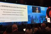
Heavy Calcification Levels PCI/CABG Playing Field at 10 Years: SYNTAXES
Calcium, one surgeon said, is a marker of a bigger problem that leads to higher mortality, regardless of treatment.

In patients who undergo revascularization, heavily calcified lesions independently predict worse survival at 10 years regardless of whether they received CABG or PCI, according to a new analysis of the SYNTAX Extended Survival Trial (SYNTAXES).
Notably, in those without heavily calcified lesions, surgery held an advantage over percutaneous revascularization in terms of mortality at 10 years, which is “consistent” with the main results of SYNTAXES, study authors, led by Hideyuki Kawashima, MD (National University of Ireland, Galway), write.
They attribute the main findings of this study both to calcium’s effects on coronary plaque morphology as well as the success of the revascularization procedures themselves. “On a macroscopic level, calcification has been linked with total plaque burden, plaque (in)stability, and delayed healing after revascularization,” Kawashima and colleagues say.
For Thomas MacGillivray, MD (Houston Methodist Hospital, TX), the study highlights “that the more you peel the onion, the more you get the sense of what a big problem coronary artery disease is. It isn't that you have it or you don't have it; there are different gradations of it,” he commented to TCTMD. “The more we study this disease, the more we learn about it and the better decisions we're able to make in terms of helping people with it.”
Importantly, MacGillivray noted, patients who have heavily calcified lesions also tend to have other risk factors. “So, it just is a marker of a bigger problem I think,” he added.
Similar Prognosis for PCI, CABG
For the study, published online last week ahead of print in JACC: Cardiovascular Interventions, Kawashima and colleagues showed that the 532 patients with at least one heavily calcified lesion in SYNTAXES had a higher crude mortality at 10 years than the 1,268 without (36.4% vs 22.3%; HR 1.79; 95% CI 1.49-2.16). Moreover, on multivariate analysis, heavily calcified lesions were an independent predictor of 10-year mortality (HR 1.36; 95% CI 1.09-1.69).
While CABG held a survival advantage over PCI among those without heavily calcified lesions (26.0% mortality at 10 years vs 18.8%; HR 1.44; 95% CI 0.97-1.41), the two procedures were associated with a similar prognosis in patients with heavily calcified lesions (10-year mortality 34.0% vs 39.0%; HR 0.85; 95% CI 0.64-1.13). These findings were confirmed on multivariate analysis.
There were also no differences in long-term mortality between PCI and CABG based upon the location of heavily calcified lesions. While patients with at least two heavily calcified lesions had higher mortality after CABG compared with those with one (44.1% vs 31.6%; HR 1.59; 95% CI 1.04-2.43), no such difference was observed in those undergoing PCI.
From a procedural standpoint, the authors explain, coronary artery calcification, for PCI, “hampers lesion crossing with stents and other devices” and also limits the ability of standard balloons to dilate stenoses optimally, often resulting in stent underexpansion and procedural complications “such as slow-flow or no-reflow, dissection, and/or perforation.” For CABG, on the other hand, the poorer survival for those with calcification could be linked with “the increased risk associated with total plaque burden and suboptimal site characteristics for graft implantation rather than impaired healing after revascularization.”
Regardless, as highly calcified lesions “reflect the aging process, complexity and extensiveness of CAD, and comorbidity, it is possible that the currently available revascularization methods do not provide benefit in the prevention of long-term mortality,” Kawashima and colleagues write. “Therefore, this study highlights the need for further research on this topic focusing on this specific population.”
Calcification as a Marker
In an accompanying editorial, Usman Baber, MD, MS (University of Oklahoma Health Sciences Center, Oklahoma City), says the lack of benefit with CABG in those with heavily calcified lesions “is somewhat unexpected and warrants further scrutiny.”
MacGillivray agreed, stressing that calcification “is a marker of a different gradation of the disease.” Perhaps, he added, CABG isn’t able to confer the same benefits as it would for those without heavy calcification, but said this is “more of a hypothesis-generating finding than a conclusion.”
MacGillivray, a surgeon, suggested that the results double down on the importance of medical management for patients with heavy calcification. “Are these patients who we should be more aggressive about treating them with higher doses of statins or different therapies in terms of anticholesterol medicines? Be more aggressive with their diabetes management? Be more focused on their medical therapies in addition to their revascularization strategies?” he questioned.
Heavily calcified lesions should be considered in the development of future revascularization risk scores, Baber suggested. The presence of highly calcified lesions, he said, may function “as an integrated imaging-based biomarker that reflects both atherosclerotic and nonatherosclerotic risk, akin to serum creatinine,” he said. “At a minimum, the inclusion of highly calcified lesions may enhance calibration of the SYNTAX score 2020, which the authors used to estimate 10-year mortality risk.”
Ultimately, this study highlights the notion of considering patient age and other comorbidities beyond coronary anatomy when deciding on how to revascularize, Baber said. “While assessing the degree of coronary complexity is the initial step in evaluating revascularization approaches, the present work reminds us we should equally consider noncoronary factors, along with patient preference, in these complex clinical decisions.”
Going forward, MacGillivray said he would like to see more attention focused on why heavy calcification puts patients at higher risk, beyond the lesions of interest. “If we can find out why the results are linked with worse survival, then we can find out how to make it better,” he concluded.
Yael L. Maxwell is Senior Medical Journalist for TCTMD and Section Editor of TCTMD's Fellows Forum. She served as the inaugural…
Read Full BioSources
Kawashima H, Serruys PW, Hara H, et al. Ten-year all-cause mortality following percutaneous or surgical revascularization in patients with heavy calcification. J Am Coll Cardiol Intv. 2021;Epub ahead of print.
Baber U. Coronary artery calcification and mortality after revascularization: look beyond the heart. J Am Coll Cardiol Intv. 2021;Epub ahead of print.
Disclosures
- The SYNTAX Extended Survival study was supported by the German Foundation of Heart Research. The SYNTAX trial, during 0- to 5-year follow-up, was funded by Boston Scientific Corporation.
- Kawashima and MacGillivray report no relevant conflicts of interest.
- Baber reports receiving honoraria/speaking fees from AstraZeneca, Biotronik, and Amgen.


Comments