Microclots Seen on OCT and Autopsy Can Inform COVID-19 Diagnostics, Care
Separate studies highlight underlying mechanisms through which the SARS-CoV-2 virus may cause heart and lung damage.

In severely ill COVID-19 patients, optimal coherence tomography (OCT) can help confirm presence of pulmonary thrombus when CT scans prove inconclusive, according to new data presented this month at TCT Connect 2020 in a special session devoted to COVID-19. A second study presented in the session added more autopsy cases to support a recent observation that some patients can have microvascular thrombi in the absence of epicardial CAD.
“We think that patients with COVID-19 pneumonia with high D-dimer levels could have lung microclots which cannot be identified by CT scan; however, they could probably benefit from anticoagulation,” said Marco Ancona, MD (San Raffaele Scientific Institute, Milan, Italy), in an email.
For the OCT study, Ancona and colleagues assessed microvascular lung thrombosis in a 58-year-old patient who presented to the emergency department with fever and cough and tested positive for COVID-19. A chest X-ray showed no signs of interstitial pneumonia, so the patient, who was diabetic and hypertensive, was discharged home on hydroxychloroquine and heparin. Six days later, he returned with difficulty breathing, at which time his chest X-ray was positive for interstitial pneumonia, so he was admitted to the ICU and placed on noninvasive ventilation. Ten days after that, the hypoxia had worsened, and his levels of D-dimer, ferritin, C-reactive protein, and interleukin-6 had all increased. A CT scan with contrast media showed ground-glass areas in the lungs, but there was no clear sign of pulmonary embolism.
Guided by published post-mortem data showing previously unsuspected segmental and unsegmental pulmonary arterial thrombosis in COVID-19 patients, Ancona and colleagues decided to perform OCT in the hopes of identifying microvascular lung thrombosis. What they found was a distal pulmonary thrombus in both lobes that appeared normal on angiography.
To TCTMD, Ancona said the patient was then treated with full dose anticoagulant therapy and had a complete recovery.
He added that the observation confirms an evolving hypothesis regarding an atypical acute respiratory distress syndrome known as MicroCLOTS—short for microvascular COVID-19 lung vessels obstructive thromboinflammatory syndrome. Ancona and colleagues are currently running an open-label, prospective study to confirm their theory in a large group of patients.
Easier Thrombus Detection
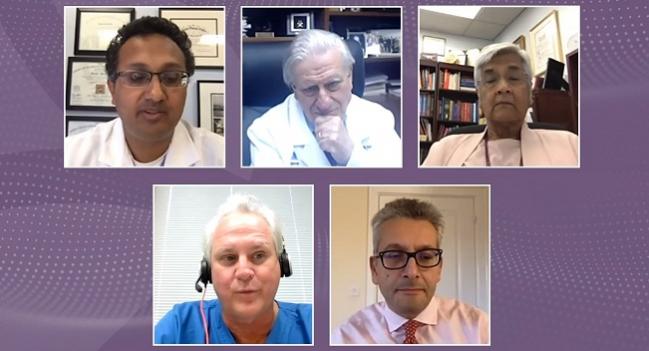
Following the presentation, panelist Renu Virmani, MD, PhD (CVPath Institute, Gaithersburg, MD), advised that OCT, rather than just CT or angiography alone, probably should be done in cases such as the one Ancona presented. “I think because there is such a high incidence . . . this is a remarkable, easy way to detect the presence of thrombus,” she said.
Moderator Timothy Henry, MD (The Christ Hospital, Cincinnati, OH), then asked what the threshold should be to use anticoagulation in these patients.
Both Virmani and panelist Valentin Fuster, MD, PhD (Icahn School of Medicine at Mount Sinai, New York, NY), said they believe that all patients hospitalized with COVID-19 should receive anticoagulation unless contraindicated. “I think it could benefit the alveolar lining as well, not just the microvasculature,” Virmani added. “The other thought is if we take away the fibrin component [through anticoagulation], we won't have so much follow-up interstitial disease.”
Multiple ongoing studies are looking at anticoagulation in hospitalized COVID-19 patients, including FREEDOM COVID-19, headed by Fuster, and IMPROVE, led by Sahil Parikh, MD (NewYork-Presbyterian/Columbia University Irving Medical Center, New York, NY), the other session moderator.
Parikh agreed that OCT may have a role “for some of these unexplained dyspneic patients who are post-COVID-19, without any sort of either macro manifestations on imaging or clinically that explain their dyspnea.”
Shedding More Light on Microthrombi
Another presentation in the same session found that microthrombi were the most frequent mechanism of myocardial necrosis in patients who died following hospitalization for COVID-19. In the study of 40 patients with confirmed myocardial necrosis, myocyte lesions were focal (< 1 cm2) in 78.6%. The data, presented by Giulio Guagliumi, MD (Ospedale Papa Giovanni XXIII, Bergamo, Italy), add more substance to a single case report recently published by his group in Circulation documenting diffuse microthrombi throughout the heart.
Virmani, who co-authored the new study, explained that she had been shocked by the amount of intramyocardial capillary thrombi when she saw that initial case report, adding: “I have never seen something like that.” On the alert for microthrombi, the investigators made a concerted effort to take as many sections from the myocardium as possible in the 40 patients, “from every wall, at two levels, so that we would not miss these things,” she said.
“When we were comparing myocardial necrosis to non-myocardial necrosis patients, epicardial coronary artery thrombus were rare and equally distributed, while microthrombi were exclusively [found] in patients with myocardial necrosis,” Guagliumi noted during his presentation.
Virmani said the data help piece together the theory that troponin elevations in COVID-19 patients may have more to do “with the fact that there is true necrosis and not so much a myocarditis. We did not see myocarditis in any of these 40 cases, except one case that had sarcoid involvement.”
L.A. McKeown is a Senior Medical Journalist for TCTMD, the Section Editor of CV Team Forum, and Senior Medical…
Read Full BioSources
Ancona M. Optical coherence tomography to assess microvascular lung thrombosis in COVID-19. Presented at: TCT 2020. October 14, 2020.
Guagliumi G. Clinical presentation and pathology of myocardial microthrombi as a cause of cardiac injury. Presented at: TCT 2020. October 14, 2020.
Disclosures
- Guagliumi, Ancona, Virmani, and Henry report no relevant conflicts of interest.
- Parikh reports institutional grant support/research contracts from Shockwave Medical, TriReme Medical, Surmodics, and Abbott Vascular; personal fees from Abiomed, and Terumo Medical Corporation; and honoraria or fees for consulting or speaking to his institution from Boston Scientific, Medtronic, CSI, and Philips.


Comments