New Chest Pain Guidelines Out at Last: Most Tests Have a Role
CT angiography gains a prominent role in the guidelines, but many other testing modalities get a class 1 indication, too.


Several years in development and beset by delays, guidelines out today from the American College of Cardiology and the American Heart Association (ACC/AHA), in conjunction with several other societies, are the first in the US addressing the evaluation and diagnosis of acute or stable chest pain.
The guidelines incorporate the use of contemporary imaging modalities, including cardiac computed tomography angiography (CCTA) and coronary artery calcium (CAC) scores, and emphasize the intensification of preventive therapies. Importantly, the guidelines also urge the more selective use of imaging and detail the factors to take into consideration when deciding between CCTA and stress testing.
“These are definitely guidelines that we felt strongly needed to be done right,” Martha Gulati, MD, chair of the guideline writing committee, told TCTMD. “Getting the data right was really important to everybody in this writing group. We had a fair number of reviewers, and we took their comments seriously. That meant that when we felt we weren’t conveying the message we wanted to make, we went back to the drawing board and asked how we could make [the guidelines] clearer and better for the practicing clinician.”
The new recommendations, published online today in Circulation and the Journal of the American College of Cardiology, are the first of their kind developed by the ACC/AHA joint committee of clinical practice guidelines.
“We weren’t updating guidelines,” said Gulati. “We didn’t have a template to go off. A lot of people seemed to mistake us for the stable ischemic heart disease guidelines, but we’re not that—we’re actually a new guideline. This is the first time the American Heart Association and American College of Cardiology wanted a guideline for the diagnosis and evaluation of chest pain.”
Ron Blankstein, MD (Brigham and Women’s Hospital, Boston, MA), one of the guideline authors, said the recommendations for cardiac testing are “revolutionary” in that they are built on a comprehensive and critical evaluation of high-quality imaging evidence.
When we felt we weren’t conveying the message we wanted to make, we went back to the drawing board. Martha Gulati
“We have more data than we’ve ever had before regarding the role of different imaging modalities,” he told TCTMD. “In the past, the role of imaging was always diagnostic, focusing on accuracy, and things like sensitivity and specificity. Now, though, the evidence base for imaging goes well beyond accuracy to include the impact of the study, cost-effectiveness, efficiency, and ultimately patient outcomes. It’s a higher quality of evidence than we’ve ever had before, which allowed us to step back and take a look at the role of imaging in different scenarios.”
For Deepak Bhatt, MD (Brigham and Women’s Hospital), another member of the writing committee, the new advice is intended to be useful to physicians on the front lines who encounter patients with chest pain, whether that’s in the emergency department or doctor’s office.
“Our goal wasn’t to focus on a procedure, or an imaging modality, or a testing modality, per se, but rather to approach it from the patient’s perspective—that being a patient who presents with a symptom—and how do we deal with that patient?” said Bhatt. “It’s not meant to replace other guidelines, such as ACS or PCI guidelines, as an example, but to integrate knowledge from a variety of different data streams, from rapidly evolving data streams, and to figure out how to provide the best care for patients with chest pain.”
’Revolutionary’ Imaging Protocols
The guidelines are endorsed by the American Society of Echocardiography (ASE), American College of Chest Physicians (CHEST), Society for Academic Emergency Medicine (SAEM), Society of Cardiovascular Computed Tomography (SCCT), and Society for Cardiovascular Magnetic Resonance (SCMR).
In broad strokes, the guidelines emphasize that “chest pain” can also mean pain, pressure, tightness, or discomfort in the chest, shoulders, arms, neck, back, upper abdomen, and jaw, as well as fatigue, shortness of breath, and nausea, particularly in women.
Early care for acute symptoms is recommended, which is an important message for the general public, while physicians are urged to follow structured risk assessment using evidence-based diagnostic protocols. High-sensitivity cardiac troponins are now the preferred standard for a biomarker diagnosis of acute MI (class 1, level of evidence B), with measurements of CK-MB or myoglobin no longer considered appropriate for the diagnosis of acute myocardial injury.
Importantly, the guideline writing committee stress the importance of identifying patients most likely to benefit from further testing, noting that more testing is not routinely needed for low-risk patients with acute or stable chest pain. For this reason, Gulati said that it’s very important to involve patients in the decision-making process, a recommendation that’s more than a throwaway line because patients are more likely to follow medical advice if they’re empowered and involved.
That’s an important part of this guidelines. Not just who to test, but potentially who not to test. Deepak Bhatt
“If we tell the patient why additional testing may not be beneficial, and why we think they’re low risk, and communicate that risk effectively to patients, they’ll be more likely to choose not to undergo additional testing,” said Gulati. “They’ll understand they’re not at risk and we’ve done what we needed to do.”
Unnecessary testing, she added, is the name of the game right now given the sheer number of Americans presenting to emergency departments or doctor’s offices with chest pain. Blankstein agreed, noting that the new guidelines emphasize that not everybody who has acute or stable chest pain—particularly low-risk patients—will require further testing.
“That’s an important part of this guideline,” added Bhatt. “Not just who to test, but potentially who not to test.”
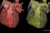
Solid Evidence Behind CCTA
When it comes down to the best way for diagnosing CAD in patients with chest pain, Gulati stressed that the appropriate test is based on the clinical scenario. There is no one test that is superior to others in all situations, she said, noting that they’ve included a number of class 1 recommendations for CCTA and functional testing.
In patients presenting to the emergency department with chest pain and suspected ACS, they advocate triaging patients into low-, intermediate-, and high-risk categories. For the intermediate-risk patient without known CAD, CCTA is now a class 1 recommendation for excluding atherosclerotic plaque and obstructive CAD (level of evidence A). If there is evidence of a stenosis, or CCTA is inconclusive, FFR-CT can be used to diagnose vessel-specific ischemia or aid clinical decisions (class 2a, level of evidence B). In the intermediate-risk patient, stress testing is also a class 1 recommendation (level of evidence B) for the diagnosis of ischemia.
Show us the trials—are they good trials, are they randomized trials, and are they comparing one type of imaging to another? Martha Gulati
For the high-risk patient with acute chest pain, or those suspected of ACS, invasive angiography is a class 1 recommendation. In the low-risk patient—those with a 30-day risk of death or MACE less than 1%—sending them home is recommended (class 2a, level of evidence B).
For the evaluation of patients with no known CAD who present with stable chest pain, again the goal is to triage patients based on risk. If low risk, CAC is considered a reasonable first-line test to exclude calcified plaque and to identify those with a low likelihood of obstructive CAD (class 2a, level of evidence B). Exercise testing without imaging is also an option to exclude myocardial ischemia (class 2a, level of evidence B). For the intermediate-to-high-risk patients with stable chest pain, CCTA is recommended to diagnose CAD, to aid in risk stratification, and to guide treatment (class 1, level of evidence A). Additionally, stress imaging with stress echocardiography, PET/SPECT, or cardiac MR can be used to diagnose myocardial ischemia and to estimate risk of MACE (class 1, level of evidence B).
“CT angiography has certainly moved up in the guidelines, but the concept of one modality moving up at the expense of another modality is not correct,” said Blankstein. “Other modalities didn’t move down because CT angiography moved up. The reality is that CT now has stronger level of evidence than other modalities. Ultimately, though, the concept is that with imaging, depending on the scenario, there are a lot of options.”
Bhatt agreed, saying there’s a role for all the different tests for the evaluation of chest pain patients.
“I wouldn’t make a blanket statement to say that this is one test that must be used in everybody,” he said. “There are circumstances that will favor the use of CT angiography. On the other hand, stress imaging might be more appropriate [in some settings].”
With multiple imaging tests now holding a class 1 recommendation, Blankstein said the guidelines were intended to be flexible, acknowledging that not every test is available at all hospitals. However, that puts the onus on physicians to select the test they think is most appropriate. The new guidelines provide some informal guidance on choosing between CCTA or stress imaging depending on the physician’s goal (for example, to rule out obstructive CAD vs ischemia-guided patient management), likelihood of obstructive CAD, prior test results, and other compelling indications.
“One message that’ll resonate with physicians practicing on the front lines is that it’s not that simple,” said Bhatt. “There are different factors that go into the assessment of the patient. Part of it is trying to understand the symptoms and that there can be overlap of different types of symptoms and overlap of different conditions. Not everything causing chest pain is due to the coronary arteries or the heart. It’s a complex evaluation.”
No Endorsement From ASNC
To TCTMD, Gulati said that they included representatives from every imaging society, including those that endorsed the new guidelines, as well as the American Society of Nuclear Cardiology (ASNC), who ultimately decided they couldn’t agree with all the recommendations and chose not to endorse the new document.
“We tried to be fair,” said Gulati. “I’m not an imager, and neither are my two co-chairs, and I think that set us up in a good way. It allowed us to ask everybody to present the most relevant evidence related to a particular imaging modality and weigh it on the same scale. Show us the trials—are they good trials, are they randomized trials, and are they comparing one type of imaging to another? Show us the data, in other words. That helped us come up with our pathways.”
Renee Bullock-Palmer, MD (Deborah Heart and Lung Center, Browns Mills, NJ), a member of the ASNC board of directors, was part of the ACC/AHA writing committee, and two ASNC members were part of the peer-review process. Despite that, the ASNC chose not to throw their support behind the recommendations.
We do appreciate the ACC/AHA trying to be collaborative, and we appreciate the complexity here. Randall Thompson
“The guidelines have been needed for a long time and it’s a complicated process, with a lot of people looking at, and having opinions on, this big body of science,” Randall Thompson, MD (Saint Luke’s Health System, Kansas City, MO), president of ASNC, told TCTMD. “In the end, we couldn’t endorse them. There were things we liked—it confirmed the importance of myocardial perfusion imaging with SPECT in several categories of patients with chest pain. It’s also nice to see other doctors recognize the value of cardiac PET. When you have an option of PET or SPECT, there are major advantages to PET.”
However, the ASNC didn’t feel the guidelines were “balanced,” said Thompson, noting that they had particular concerns about the use of FFR-CT.
“In our opinion, it wasn’t ready for the prominent role it was given,” he said, adding that the guidelines fail to sufficiently document the shortcomings of the new technology. In several clinical scenarios, FFR-CT or stress imaging can be used as an add-on test to aid in decision making and diagnosis, such as in intermediate-risk patients with acute or stable chest pain and no known CAD. For Thompson, the new guidelines also tend to overemphasize CCTA while blurring the distinction between the different stress testing modalities.
Despite giving their feedback, Thompson said they couldn’t get the ACC/AHA writing committee to alter the recommendations based on their concerns, which led them not to endorse the chest pain guidelines. “We do appreciate the ACC/AHA trying to be collaborative, and we appreciate the complexity here, too,” said Thompson. “There is a need to compromise a little bit, but this was just too far. It wasn’t patient first, and it wasn’t the right test in the right patient.”
Not Just ‘Chest’ Pain
Language also gets a tweak in the new guidelines, with “atypical” chest pain out as a descriptor. Instead, the guideline authors recommend that “noncardiac” chest pain be used if heart disease is not suspected.
“I’ve never been a fan of it,” said Gulati. “It’s a label used more frequently with women, but atypical, by definition, means presenting a little bit differently, and the problem is that it’s been misinterpreted. When my fellows tell me that this is a person with atypical chest pain, they’re trying to convey that they don’t think it’s cardiac. I think the term has been misused.”
CT angiography has certainly moved up in the guidelines, but the concept of one modality moving up at the expense of another modality is not correct. Ron Blankstein
Atypical is a commonly used term, said Bhatt, but it can lead to misdiagnosis and undertreatment in women.
“There are a variety of noncardiac symptoms that can occur that represent ischemic coronary manifestations,” he said. The majority of men and women with significant CAD present with chest pain or pressure, Bhatt added, in addition to other noncardiac symptoms. “Often when we’re hearing fatigue or dyspnea, there is some chest pain, discomfort, or pressure also mentioned.”
Blankstein said the new guidelines now characterize nonobstructive CAD, which is often defined as luminal stenosis < 50%, as “having known CAD,” which has implications for further testing or aggressive prevention. The guidelines, he said, provide multiple opportunities in the stable and acute chest pain patients for physicians to defer further testing and to optimize medical therapy.
Blankstein noted that one additional way the guidelines attempt to minimize unnecessary testing is by highlighting that prior cardiac testing carries with it a “warranty” period in patients with chest pain. The warranty period for a normal coronary angiogram or CCTA with no stenosis or plaque is 2 years, while a normal stress test is good for 1 year.

Michael O’Riordan is the Managing Editor for TCTMD. He completed his undergraduate degrees at Queen’s University in Kingston, ON, and…
Read Full BioSources
Gulati M, Levy PD, Mukherjee D, et al. 2021 AHA/ACC/ASE/CHEST/SAEM/SCCT/SCMR guideline for the evaluation and diagnosis of chest pain: a report of the American College of Cardiology/American Heart Association joint committee of clinical practice guidelines. Circulation 2021;Epub ahead of print.
Disclosures
- Gulati reports no conflicts of interest.
- Blankstein reports research funding from Amgen Inc and Novartis; serving on the steering committee for Vesalius trial (Amgen); consulting for Caristo Diagnostics Inc.
Related News


Comments