What’s the Damage With Long COVID? Advanced Imaging Reveals Clues
Researchers used PET/MRI to detect otherwise invisible inflammation. Less clear is how this might influence patient care.

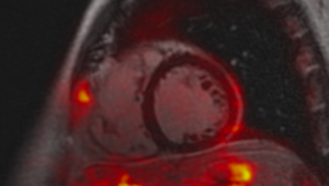
Photo Credit: Maria Giovanna Trivieri
Many patients referred for specialty care to address long-COVID symptoms have subtle evidence of inflammation and other cardiopulmonary abnormalities that can be detected with advanced imaging, a single-center study shows.
Researchers performed 18F-FDG PET/MRI of the heart and nearby blood vessels as well as dual-energy CT of the lungs and analysis of plasma protein levels to pinpoint these features about a year after SARS-CoV-2 infection. They also kept tabs on patients’ clinical outcomes for nearly 4 years.
“What this study shows is that there are some hidden changes caused by COVID that are apparent when you use sophisticated imaging,” said Maria Giovanna Trivieri, MD, PhD, who led the study along with Ana Devesa, MD, PhD (both from Icahn School of Medicine at Mount Sinai, New York, NY).
More typically, patients undergo screening with more common tools like echocardiography, MRI, or CT, she said. But for some individuals, “if you don’t use the right tool, you might not see what you’re interested in identifying.”
Colin Berry, MBChB, PhD (University of Glasgow, Scotland), pointed out that “PET/MRI is very much a research tool,” one that is primarily available at expert centers and involves exposure to ionizing radiation. Thus, the study results might not apply to clinical practice on a large scale, he told TCTMD.
Moreover, these long-COVID patients were not “all-comers” but rather were sick enough to merit extra scrutiny. “We wouldn’t wish for this to trigger, globally, people with long COVID [saying] they need to have a PET/MRI scan. I don’t think that’s really at all the conclusion here,” Berry said, adding that there could be a role for such imaging in selected cases.
More than 5 years after the pandemic began, it’s also becoming clearer that at least some patients will see their long-COVID symptoms resolve over time, Berry noted. “In general, there’s an improving trajectory.” Additionally, many—though not all—people with the condition had preexisting health problems, which makes it hard to tease out what’s causing their breathlessness, fatigue, and other difficulties.
According to Berry, the best path forward in this field may lie in better understanding self-management rather than a sophisticated cure. “The reason I say that is because the large majority of people with long COVID are in the community. They’re attending primary care,” he said. “Their symptoms might extend across systems: respiratory, cardiovascular, GI. The idea that one medicine is going to improve that—in other words, [that] we need to wait for drug trials—that’s never going to happen, so we need to identify nonpharmacological therapies that lead to patient benefit.”
Such therapies could relate to mental health, overall well-being, physical activity, and the like, he suggested, adding that educational materials to empower patients in these areas do exist but need to be more widely available.
PET/MRI, Dual-Energy CT, and More
For the study, published recently in the Journal of Nuclear Medicine, Trivieri, Devesa, and colleagues enrolled 98 patients (median age 48.5 years; 53% women) with lingering symptoms 9 to 12 months after a bout of COVID-19. Four had a history of heart disease, all of whom had heart failure (three with preserved ejection fraction and one with reduced ejection fraction). All had normal brain natriuretic peptide levels when enrolled in the study.
Long-COVID symptoms included shortness of breath (80%) as well as fatigue, myalgias, fever, and chills. Nearly three in 10 were hospitalized at the time of the study.
PET/MRI scans were done at a median of 303 days after the initial onset of COVID-19. More than half (57%) had abnormal findings: 24% had signs of myocarditis, 22% had signs of pericardial involvement, 11% had periannular uptake, and 30% had vascular uptake (aortic or pulmonary).
The researchers also compared these individuals with nine controls (mean age 42 years; 56% women). On PET/MRI, none had signs of myocardial involvement, pericardial involvement, or periannular uptake.
Blood samples for 24 of the long-COVID patients were taken around the time of PET/MRI. Interleukin-7, -2, -10, and -17A levels all were elevated in the long-COVID versus control group, while levels of interferon-γ, tumor necrosis factor, and vascular endothelial growth factor were decreased. Biomarkers also varied within the long-COVID group between patients with abnormal versus normal PET/MRI results.
Among the 70 patients who underwent dual-energy CT, 67% had pulmonary infiltrates and 59% had abnormal perfusion.
During a median follow-up of 3.8 years, nine of the long-COVID patients developed heart failure (four with preserved and five with reduced ejection fraction), two developed mitral valve disease, and one developed another valvular disease. Myocardial infarction, pulmonary embolism, and supraventricular tachycardia occurred in one patient each. Additionally, one of the patients who had heart failure with reduced ejection fraction developed secondary pulmonary hypertension.
Two-thirds of patients who experienced cardiac events had abnormal PET/MRI scans. The proportion was even higher—78%—among those who eventually had heart failure. However, the associations between abnormal imaging and subsequent cardiac events or heart failure did not reach statistical significance.
Patients with abnormalities on imaging or in their biomarkers “might warrant further monitoring to exclude the development of complications such as pulmonary hypertension and valvular disease,” the researchers say, though they caution that they “cannot confidently rule out that some of these abnormalities were preexisting.”
Overall, their study results “suggest different pathologic mechanisms or etiologies and may foreshadow the development of future cardiovascular morbidity, suggesting that COVID infection should be considered a risk factor for future cardiopulmonary sequelae,” they conclude.

Caitlin E. Cox is Executive Editor of TCTMD and Associate Director, Editorial Content at the Cardiovascular Research Foundation. She produces the…
Read Full BioSources
Trivieri MG, Devesa A, Robson PM, et al. Prevalence of persistent cardiovascular and pulmonary abnormalities on PET/MRI and DECT imaging in long COVID patients. J Nucl Med. 2025;Epub ahead of print.
Disclosures
- The study has been partially supported by the Thrasher Research Fund and the Spanish Society of Cardiology.
- Trivieri was supported by grants from the National Institutes of Health and the American Heart Association.
- Devesa is recipient of the Alfonso Martin Escudero grant.
- Berry reports no relevant conflicts of interest.
Related News


Comments