CCTA Increases Contrast Volume in Patients With Prior CABG: GREECE Trial
A bigger trial with newer scanners is the next step in learning how to better assess these challenging patients, a researcher says.

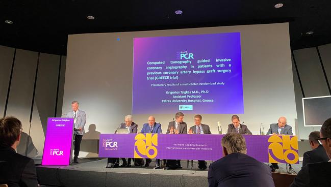
PARIS, France—On top of invasive coronary angiography alone, performing coronary CT angiography (CCTA) first to evaluate hemodynamically stable patients with prior CABG does not lower but rather increases the amount of contrast used, albeit without spurring more contrast-induced nephropathy, according to data from the randomized GREECE trial.
While GREECE failed to meet its primary endpoint, Grigorios Tsigkas, MD, PhD (Patras University Hospital, Greece), who presented the findings last week at EuroPCR 2022, said use of CCTA for these complex patients remains promising given that it lowered procedural times by enabling quicker decision-making and that it decreased the risk of MACE at both 5 and 30 days.
“Coronary angiography post-CABG is always a challenge,” he said. “It is obvious that we need a bigger trial with probably newer CT scanners, and that could lead to better results, focusing mostly on radiation. As we manage true complex patients, a real scenario—and very feasible to do—is to use better technology with [a] final result [leading] to less contrast, less radiation, and safe procedures in less time.”
Discussing the results following the presentation, co-moderator Chaim Lotan, MD (Hadassah-Hebrew University, Jerusalem, Israel), said interventional cardiologists still have much to learn about interpreting CT results. “It's not so obvious for all of us how do we analyze and how do we get into the valve,” he said. “Once we have more experience, it should enhance the way that we find the venous grafts—[saving] more time and contrast and everything.”
More Contrast, Less Time, Fewer MACE
For the study, Tsigkas and colleagues randomized 153 patients (mean age 70 years; 82% male) with prior CABG and a clinical indication for coronary angiography to invasive angiography alone (n = 69) or to CCTA with about 90 mL of contrast agent followed by invasive angiography (n = 84). Almost all patients (87.6%) had a known surgical report, just under half had a prior MI, slightly more than half had stable CAD, and just over one-third had prior PCI.
The volume of contrast agent administered (primary endpoint) was significantly greater in the patients who first underwent CCTA, but the number of catheters used and the total procedure time were lower compared with controls. Additionally, total effective radiation dose was higher in the study group.
Procedural Results
|
|
CCTA + (n = 84) |
Invasive (n = 69) |
P Value |
|
Total Contrast Volume, mL |
209.1 |
165.3 |
0.006 |
|
Total Contrast Volume |
175.7 |
125.3 |
< 0.001 |
|
Total Catheters Used |
2.8 |
3.2 |
0.049 |
|
Procedural Time, min |
28.5 |
38.4 |
0.022 |
|
Total Effective |
24.66 |
11.91 |
< 0.001 |
Also commenting on the findings, panelist Petr Widimský, MD, DrSc (Charles University, Prague, Czech Republic), said the study may have been more successful if it had made invasive coronary angiography optional instead of mandatory in the study arm. “In such a strategy, you could end up with [a] major part of the first group with only CT angiography,” he said.There were no significant differences in postprocedural complications within 6 hours (7.1% vs 2.9%; P = 0.295) or contrast-induced nephropathy (16.0% vs 13.8%; P = 0.714) between the study and control groups. However, MACE rates within 3 to 5 days (0 vs 11.6%; P = 0.001) and after 30 days (5% vs 16%; P = 0.023) each were lower in the CCTA arm.
Tsigkas said this was a purposeful design. “There was initially a clinical indication for coronary angiography, so we used the information we took from CT angiography and after that we went directly to compare the results,” he explained. “Our initial thought was to use less contrast and less time to check the native coronary arteries [with invasive coronary angiography].”
Further, Tsigkas added, “real-life evidence shows that among all-comers patients after CABG, about 30% go directly for the PCI. In other words, initially a conservative management is the first suggestion, I would say.”
“CT is at the first stages, and it's going to impact significantly,” Lotan concluded. “In a few years, once each one of us [has] more experience with CT analysis, I think it will be different, and I agree with Dr. Widimský that maybe in some cases, we don't have to intervene. The oculostenotic reflex that we have in angiography may be a little bit changed when we have CT, and especially in the future where we have CT FFR, [we’ll] know whether we have to do anything or not at all.”
Yael L. Maxwell is Senior Medical Journalist for TCTMD and Section Editor of TCTMD's Fellows Forum. She served as the inaugural…
Read Full BioSources
Tsigkas G. Computed tomography guided invasive coronary angiography in patients with a previous coronary artery bypass graft surgery trial (GREECE trial). Presented at: EuroPCR 2022. May 17, 2022. Paris, France.
Disclosures
- Tsigkas, Lotan, and Widimský report no relevant conflicts of interest.


Comments