DISRUPT PAD III: Lithotripsy Shows Promise in Heavily Calcified PAD
Delivery of sonic pressure waves was associated with fewer severe dissections, postdilations, and bailout stents than PTA.

A catheter-based device that uses shockwaves to crack calcium in femoropopliteal arteries appears safe and effective in the short term, with fewer dissections as well as less use of postdilation and bailout stenting compared with percutaneous transluminal angioplasty (PTA), according to data from the DISRUPT PAD III trial.
“[Intravascular lithotripsy] was superior to PTA in acute procedural success and demonstrated atraumatic treatment,” said William A. Gray, MD (Mainline Health, Wynnewood, PA), presenting the RCT results this Saturday at VIVA 2020. The findings are in line with those from observational registries where the device (Shockwave Medical) was used in multiple vessel beds, he added.
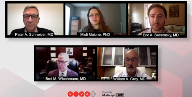
Following the presentation, moderator Peter Schneider, MD (University of California, San Francisco), asked Gray if he would consider the device as a stand-alone therapy.
“It’s really enabling an adjunctive technology. It gets you through the procedure, gets a better result with lower complication rates,” Gray replied. “I don't believe anybody thinks it's going to be a stand-alone procedure as relates to tangible evidence.” However, he noted that the majority of patients did not require a stent or postdilation following lithotripsy.
Percent Diameter Stenosis Goal Achieved
For DISRUPT PAD III, 306 patients from 45 centers in the United States, New Zealand, Germany, and Austria were randomized to intravascular lithotripsy or PTA, followed by additional treatment with a drug-coated balloon (DCB) or stent if needed. The majority of patients in each treatment group were Rutherford category 3, with no differences in lesion length, calcified length, or severity of calcification.
Fluoroscopy times averaged 3 minutes longer in the lithotripsy group, which Gray attributed to older generators not allowing for more than 180 pressure-wave pulses per balloon—the average number of pulses needed was 228. The current generator, he added, allows up to 300 pulses, which should reduce fluoroscopy times. Approximately 5% of patients required postdilation in the lithotripsy group, compared with 17% in the PTA group (P = 0.001). Similarly, the rate of stent placement was 4.6% in the lithotripsy group versus 18.3% in the PTA group (P = 0.0002).
Following the procedure, 66% of the lithotripsy patients achieved the goal of angiographic core lab-confirmed diameter stenosis ≤ 30% (without flow-limiting dissection and prior to DCB +/- stenting), compared with 50% of the PTA group (P = 0.02). Site-reported success was even higher, with 90% of lithotripsy patients and 65% of PTA patients having met the ≤ 30% residual stenosis benchmark.
Final percent diameter stenosis was 27.3% with lithotripsy versus 30.5% with PTA (P = 0.04).
Additionally, angiographic complications were minimal with lithotripsy, with an 81.5% freedom from any dissection; it was 67.7% in the PTA arm. For dissections equal to or greater than grade C, those rates were 3.5% and 15.1%, respectively—a 77% relative risk reduction, Gray noted.
For the primary endpoint of residual stenosis ≤ 30 percent without flow-limiting dissection grade D or worse prior to DCB, success was reported in 90.1% of the lithotripsy group and 64.5% of the PTA group (P < 0.0001). On core-lab adjudication, the difference between groups remained at over 15%.
Final angiographic and clinical outcomes showed that after the allowance of further dilation and stent implantation, both groups had similar reference vessel diameters, minimum lumen diameters, diameter stenoses, acute gains, dissections, ankle-brachial indexes, and walking impairment scores, as well as similar improvements in Rutherford category.
In terms of safety, lithotripsy was not associated with major adverse outcomes, emergency revascularization, major amputation, thrombus or distal embolization, or perforation. At 30 days, the rate of clinically driven TLR was 0.7% in both treatment groups.
Another Tool for Complex Patients
Eric Secemsky, MD (Beth Israel Deaconess Medical Center, Boston, MA), one of the panelists for the session, has used the device in both peripheral and coronary procedures. To TCTMD, he said the ease of use and safety are important, although long lesions may be less suitable for this type of therapy.
“Where I see value of this is in areas where I do not want to stent,” he said. “There also could be some role in in-stent restenosis and other types of failed prior endovascular lesions that require more-aggressive therapy, but we don't have a lot of indicated devices for those segments.”
During the session, Secemsky asked Gray whether there were differences in how the device worked in patients with different types of calcium. “I don't know that there's a difference. In my experience with the eccentric calcium, it seems to work just as well as it does with the concentric calcium,” Gray said.
It gives you a nice tool to safely address particularly complex lesions and particularly complex territories. Eric Secemsky
The study results come on the heels of the recent presentation and publication of the nonrandomized DISRUPT CAD III trial, in which intravascular lithotripsy was shown to help optimize PCI in severely calcified coronary lesions.
“I think the key for lithotripsy in the lower extremities is a little bit different than in the coronaries. We're not only looking for good lesion expansion and potentially for optimizing lesions to be adjunctively treated with drug-coated therapies, such as drug-coated balloon angioplasty, but we're also looking for safety in certain areas where we don't want to have to be concerned about bailout stenting,” Secemsky said. “It gives you a nice tool to safely address particularly complex lesions and particularly complex territories.”
L.A. McKeown is a Senior Medical Journalist for TCTMD, the Section Editor of CV Team Forum, and Senior Medical…
Read Full BioSources
Gray WA. IVL for peripheral artery calcium: the DISRUPT PAD III randomized controlled trial 30-day outcomes. Presented at: VIVA 2020. November 7, 2020.
Disclosures
- Gray reports consulting, and research, clinical trial, or drug study funding from Shockwave Medical.
- Secemsky reports speaking and consulting fees from Abbott, BD, Bayer, Cook, CSI, Janssen, Medtronic, and Philips; and research grants to his institution from AstraZeneca, BD, Boston Scientific, Cook, CSI, Medtronic, and Philips.


Comments