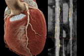
Fewer Women Than Men Get Intracoronary Imaging in PCI, but Why?
A British registry shows a widening sex gap. More worrisome: the data also link imaging to better survival.


Women undergoing PCI in England and Wales are less likely to receive intracoronary imaging than their male counterparts, data from the British Cardiovascular Intervention Society (BCIS) registry show. The situation grew worse, not better, over a 14-year period.
Importantly, female patients may be paying a penalty, too, in terms of survival and major adverse outcomes, Muhammad Rashid, MBBS (Keele University, Stoke-on-Trent, England), and colleagues point out in a new paper, which was published online earlier this week in JACC: Cardiovascular Interventions.
Rashid told TCTMD that their study comes on the heels of others showing women have worse outcomes when they undergo PCI. But whether that’s due to biological or clinical factors, or to differences in care, isn’t known. “Intracoronary imaging is obviously one of the important modalities which help to optimize procedures,” with the goal of improving outcomes, he said, and they wanted to figure out whether this might explain some of the disparities tied to sex.
It’s particularly concerning, Rashid said, that the gap in use of IVUS and optical coherence tomography (OCT) between men and women continued to grow, reaching an absolute difference of 5% by the end of the study period.
“What I was hoping actually was that over time, as the use of imaging increased, we would see more equity,” he continued. Instead, the curves diverged. “Initially we saw that there was almost no difference in uptake of intracoronary imaging in both men and women, but as the overall number of intracoronary imaging[-guided] PCIs increased in UK practice, we began to see a divergence in the curves . . . over the last 5 years.”
Michelle Williams, MBChB, PhD (University of Edinburgh, Scotland, and British Heart Foundation Data Science Centre), president elect of the British Society of Cardiovascular Imaging/British Society of Cardiovascular CT (BSCI/BSCT), also pointed to this difference as striking, given that it persisted even as the knowledge base grew.
Speaking with TCTMD, Williams said the findings confirm what she and others found when analyzing BSCI/BSCT data. Their study, which focused on an “earlier stage in the pathway,” showed “women are less likely to get invasive investigation after an abnormal CT scan,” she noted. This new paper “really nicely shows that even once they get the invasive investigation, they’re less likely to get the intracoronary imaging to appropriately guide revascularization.”
All PCIs in England and Wales
Using the BCIS registry, Rashid et al analyzed data on nearly a million adults who underwent PCI in England and Wales between 2006 and 2019. Intracoronary imaging (intravascular ultrasound and optical coherence tomography) was used in 8.4% of the 738,616 men and 7.9% of the 255,862 women (P < 0.001).
There are still lots of questions about what it actually means in clinical practice, but it’s really important that we’ve seen this. The next question is: what are we going to do about it?” Michelle Williams
Compared with women who didn’t receive intracoronary imaging, those who did were more likely to belong to an ethnic minority group (17.1% vs 13.7%), to have left main disease (22.6% vs 3.0%), and to be undergoing multivessel PCI (34.5% vs 18.6%), with a greater likelihood of being treated for three or more lesions (12.0% vs 6.8%).
Intracoronary imaging became more widely adopted over the years, but by the end of the study period just 14.5% of women received it during PCI, as compared with 19.6% of men (P < 0.001).
Female sex, after accounting for potential confounders, independently predicted lower use of intracoronary imaging (OR 0.93; 95% CI 0.91-0.96). Women received less imaging than men irrespective of disease complexity, with consistently lower rates whether they presented with ACS, long lesions, chronic total occlusions, left main disease, in-stent restenosis, stent thrombosis, calcified lesions, or renal disease.
Yet, after accounting for clinical and angiographic differences, both sexes saw better survival and fewer MACCE with intracoronary imaging, albeit accompanied by a higher risk of bleeding.
Outcomes With vs Without Intracoronary Imaging: Adjusted OR (95% CI)
|
|
Men |
Women |
|
In-Hospital Mortality |
0.48 (0.44-0.53) |
0.56 (0.48-0.64) |
|
In-Hospital MACCE |
0.75 (0.71-0.80) |
0.83 (0.76-0.91) |
|
Bleeding Complications |
1.19 (1.03-1.37) |
1.28 (1.09-1.50) |
The results support the notion that intracoronary imaging can improve outcomes by characterizing plaque morphology and optimizing stenting, the researchers say. However, they acknowledge, the registry lacks details on sex-related anatomical differences and how the information derived by imaging actually was applied to optimize stent placement and deployment.
This study valuably demonstrates how large-scale healthcare data can provide key insights, said Williams. “If you’re just dealing with one center, looking at the data from one place, you wouldn’t really get [the entire] picture. Whereas this [captures all of] England and Wales, and we know that there is a systematic issue across the board.”
Future studies should look into whether these patterns exist elsewhere in the world, she added.
And while imaging use was associated with superior outcomes, what’s not known from the current study is what the imaging—when it was done—specifically showed, Williams observed. “So, what did it find and how did the findings translate to [decision-making] and impact the outcomes? There are still lots of questions about what it actually means in clinical practice, but it’s really important that we’ve seen this. The next question is: what are we going to do about it?”
Potential reasons women receive less intracoronary imaging include having smaller arteries or different disease distribution compared with men, Williams suggested, or it could be that clinicians may simply not be treating female and male patients the same.
For Rashid, another possible explanation for the sex gap is that many women have atypical presentations. Regardless, the exact reasons need to be established, he noted. “We need to accept that there is a disparity and then we need to devise strategies to try and improve that [to] offer better care to our patients.”
These data are observational, Rashid acknowledged, so moving forward, it’s important to enroll more women in RCTs of intracoronary imaging. In some ways, such imaging might prove to be particularly useful in female patients, who experience a higher incidence of myocardial infarction with nonobstructive coronary disease, spontaneous coronary artery dissection, and plaque erosion. “Imaging really helps to define, first of all, the diagnosis and, more importantly, [helps] to tailor treatment,” he said.
At the very least, Williams said, imaging is “just as useful” in women as in men, if not more so.
Clinicians need to be aware that “women present with coronary artery disease just the same as men,” she stressed. “We need to be thinking about how we’re using things appropriately and equally, and not missing things just because someone is a woman or a man.”

Caitlin E. Cox is Executive Editor of TCTMD and Associate Director, Editorial Content at the Cardiovascular Research Foundation. She produces the…
Read Full BioSources
Rashid M, Stevens C, Zaman S, et al. Sex differences in use of intracoronary imaging in patients undergoing percutaneous coronary intervention. J Am Coll Cardiol Intv. 2022;15:1283-1295.
Disclosures
- Rashid and Williams report no relevant conflicts of interest.
Related News


Comments