Radiation Exposure ‘Extraordinarily’ High for Imagers in Structural Procedures
Experts call for greater awareness and standardization of proper protection methods in cath labs as this field grows.

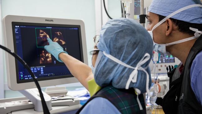
Photo Credit: Rebecca Hahn
Compared with interventional cardiologists, interventional imagers are exposed to markedly higher levels of radiation during structural heart procedures, according to new single-center data. These levels range from threefold higher for left atrial appendage occlusion (LAAO) procedures to 12-fold higher for transcatheter edge-to-edge repair (TEER) of mitral valve disease, investigators say.
“We did hypothesize that interventional echocardiographers were going to have higher radiation doses,” because of their positioning closer to the source of radiation, lead author David A. McNamara, MD (Spectrum Health, Grand Rapids, MI), told TCTMD. “However, the degree to which we saw this was greater than we expected.”
The study is not the first to draw attention to this cath lab occupational hazard, with a 2018 paper showing that ceiling-mounted lead shielding can reduce radiation exposure for transesophageal echocardiography (TEE) operators by as much as 82%.
Vast Differences
For their study, published online last week in JAMA Network Open, McNamara and colleagues collected radiation data from interventional echocardiographers, interventional cardiologists, and sonographers from their institution who performed 30 sequential LAAO and 30 sequential TEER procedures between July 2016 and January 2018.
Mean age was 79 years, 53.3% of patients were male, and all were considered to be at high cardiovascular risk. Median fluoroscopy time was longer for TEER than for LAAO procedures (20.9 vs 9.2 minutes; P < 0.001).
We hope that this shines a light on the importance of recognizing occupational radiation exposure and helps promote further radiation mitigation research. David McNamara
Interventional echocardiographers were exposed to an overall higher median radiation dose per case than interventional cardiologists (10.6 vs 2.1 μSv). This finding was maintained when looking individually at TEER (10.5 vs 0.9 μSv) and LAAO (10.6 vs 3.5 μSv; P < 0.001 for all).
Sonographers reported lower median radiation doses than interventional imagers during both LAAO (0.2 μSv) and TEER procedures (0.0 μSv; P < 0.001 for both).
At his hospital, McNamara said, they have “a high degree of radiation safety embedded in its structure.” Notably, they use “quite a bit more shielding and more lead protection than do other institutions at which I've trained and been in communication with,” he noted. “We strongly believe that this is not an issue with the institution that this was conducted, this is really more of a highlighting this as a field is something that we need to be more cognizant of.”
He said he hopes these findings will increase awareness of the sometimes dangerous levels of radiation that are “frankly underappreciated right now by those that perform these procedures. . . . We hope that this shines a light on the importance of recognizing occupational radiation exposure and helps promote further radiation mitigation research.”
Different Setup Options
Commenting on the study for TCTMD, Dee Dee Wang, MD (Henry Ford Hospital, Detroit, MI), said data like these are important to codify the emerging subspecialty of interventional imaging, especially since these physicians are often “taken for granted.”
“If you think about it from a philosophical standpoint, you get to have tenfold the radiation, but you get to have one quarter of the RVU productivity,” she said. “This is important to emphasize that this is a new subspecialty within cardiology. It's got its own procedural risks, just like interventional cardiology.”
You want to have longevity this career, you want to continue to be able to do what you love, but nobody wants a cataract in their early forties. Dee Dee Wang
Those who have chosen the career path of helping guide these procedures obviously are aware of some degree of radiation exposure, but they “absolutely” don’t recognize the current disparities at most cath labs around the country, Wang continued.
“Not everybody is able to advocate or understand what they need, and hopefully from this paper, they should understand the bare minimum and what they must request before going to a room for their own safety,” she stressed, adding, “They need to be aware of what's going on and how they need to protect themselves and advocate for their safety. You want to have longevity this career, you want to continue to be able to do what you love, but nobody wants a cataract in their early forties.”
Both Wang and Rebecca Hahn, MD (NewYork-Presbyterian/Columbia University Irving Medical Center, New York, NY), told TCTMD their current protection setups are different from what was documented in the study in that they always use mounted floor-to-ceiling shields. Additionally, Hahn said she no longer stands next to the head of the patient, but rather to the side with a rolling shield to protect her head and neck. Hahn said she would be “surprised if I had higher numbers than our interventionalists.”
Likewise, because of these protective measures, Wang said her radiation exposure is “much, much less than what was documented in this paper.”
Even so, Hahn said she “love[s] the fact that they're bringing attention to the fact that greater protection needs to be performed. But even more important than that, . . . [many] have yet to accept the fact that interventional imaging is actually a separate subspecialty that requires some guidelines. Right now we just have some consensus papers, . . . and so I like that that they tried to highlight the fact that we are extraordinarily exposed.”
‘A Team Sport’
Hahn called structural heart interventions “a team sport” and acknowledged that most interventional cardiologists “are very conscious of [not] exposing the imager to more radiation than we have to have.” When Hahn has to move her probe, she alerts her colleagues so that they temporarily pause fluoroscopy. “There's no reason for them to fluoro me while I'm changing the image. They don't need to watch my hands moving,” she said.
Yet even with conscientious colleagues and high-quality equipment, other considerations need to be made to increase operator safety. “The machine settings for voltage and charge and frame rate will play a big role, but [reducing] distance is one of the best ways of reducing radiation because it drops off exponentially your exposure,” Hahn said, adding that she trains fellows to hold the long, stiff TEE probes down away from the patient’s mouth so as to reduce exposure to the hands. She also called for lead skirts to be installed on both sides of the operating table as well as greater use of rolling shields or even mobile chariots that “wrap around the imager.”
We are extraordinarily exposed. Rebecca Hahn
Wang also called for increased use of CT preprocedural planning. “For our left atrial appendage procedures, we published that we can decrease the procedure time by at least 10 to 15 minutes, sometimes as much as 30 minutes, because we know what device size to use,” she said.
While no cardiovascular or imaging societies have yet endorsed any kind of standardized occupational safety procedures for interventional imaging physicians, Wang said this paper, even though from a single-center, “is a great call to action now that we have data. . . . I don't think you need to do a multicenter study to actually prove anything more beyond what two peer-reviewed manuscripts have documented in a span of 4 years.”
Standardization will become even more important as the field of structural heart interventions grows. Tricuspid interventions can last up to 6 hours, Wang highlighted. “That's a ton of more radiation exposure than you would have for MitraClip procedure.”

Yael L. Maxwell is Senior Medical Journalist for TCTMD and Section Editor of TCTMD's Fellows Forum. She served as the inaugural…
Read Full BioSources
McNamara DA, Chopra R, Decker JM, et al. Comparison of radiation exposure among interventional echocardiographers, interventional cardiologists, and sonographers during percutaneous structural heart interventions. JAMA Network Open. 2022;5(7):e2220597.
Disclosures
- McNamara, Wang, and Hahn report no relevant conflicts of interest.
Related News


Comments