Sodium Fluoride Imaging in AAAs Provides a Picture of Future Risk
The PET/CET approach seems adept at identifying which AAAs are likely to grow, rupture, or require repair, researchers say.

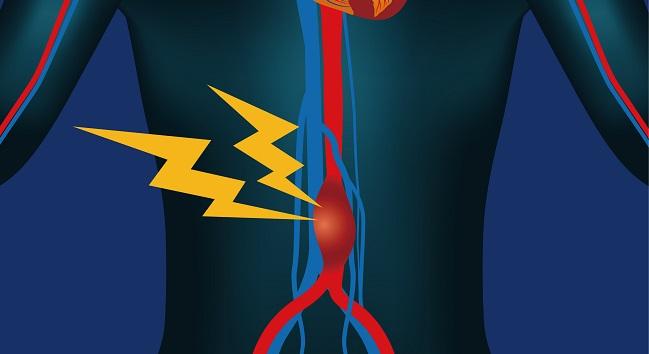
In patients with abdominal aortic aneurysm (AAA), identifying areas of sodium fluoride uptake on PET and CT can help pinpoint which regions in the aorta are likely to grow over time and which individuals are likely to one day have clinical consequences, according to early data on a novel approach.
Specifically, the SoFIA3 trial found that patients whose aneurysms were in the highest tertile of uptake saw their aneurysms grow more than twice as quickly as those in the lowest tertile and were nearly three times more likely to experience AAA rupture or require repair.
“This is the first study to demonstrate that an imaging biomarker of disease activity can add to the risk prediction of AAA and to suggest that this approach might refine clinical decisions regarding the need for surgery and improve patient outcomes,” Rachael O. Forsythe, MD (University of Edinburgh, Scotland), and colleagues report in their paper published in the February 6, 2018, issue of the Journal of the American College of Cardiology.
Since AAAs that grow over time are vulnerable to rupture, which can often be fatal, patients with the condition typically are tracked with ultrasound. However, as Forsythe and colleagues point out, “AAA growth is nonlinear, unpredictable, and influenced by biomechanical processes that cannot be predicted by conventional anatomic imaging alone. Indeed, aneurysms not infrequently rupture below the current threshold (55 mm in diameter) for elective repair, and many patients with aneurysms with > 70 mm never experience rupture.”
To meet this clinical need, the researchers looked into a possible early-warning system: sodium fluoride uptake on PET/CT, which has been shown in carotid and coronary atherosclerotic plaques to identify early microcalcification that occurs in response to inflammation. This biological activity, they say, may indicate which AAAs are likely to cause eventual harm.
If you can get at the process before it’s going to manifest in something catastrophic in the future and intervene at that stage, you can apply that to many [diseases]. I think that’s a very attractive prospect. Rachael O. Forsythe
“The radiotracer we’ve used and the PET/CT technique is something that’s been used for 40 years, so we knew it was very safe,” Forsythe told TCTMD. “It’s readily available in centers that can do PET/CT and can manage the tracer. So, from the technology point of view, we’re already there.” While not every hospital has this technology, she added, “it’s becoming more widespread.”
Specific to AAA, the real-world use of the approach explored in SoFIA3 requires further study, but the potential benefit to patients is large, Forsythe said. “It’s a disease process with very high mortality if left untreated, and if an aneurysm ruptures, the overall mortality is up to 90% and that includes people who get operated on.”
The key question, she said, about measuring sodium fluoride uptake on PET/CT is: “Is it necessary over and above what we do?” Ultrasound is cheaper and doesn’t involve any radiation, Forsythe observed. “But what our study has shown is that this technique adds value to assessing aneurysms beyond ultrasound.”
AAA Growth and Rupture
For the SoFIA3 trial, which included a prospective case-control study as well as a longitudinal cohort study, participants underwent PET/CT imaging to assess fluorine-18-sodium fluoride (18F-NaF) uptake as well as abdominal ultrasound, CT angiography, and calcium scoring.
In the case-control study, 20 patients with AAA were matched to 20 control subjects based on age, sex, and smoking status. Uptake of 18F-NaF was higher in the AAAs than in the normal abdominal aortas of the controls (P = 0.023). Additionally, “histology and micro-PET-CT demonstrated that 18F-NaF uptake localized to areas of aneurysm disease and active calcification,” the investigators report.
The longitudinal cohort study involved 72 patients with AAA who had a mean age of 73 years and a mean baseline aneurysm diameter of 48.8 mm. Within the AAAs of those individuals, 18F-NaF uptake was higher than in nonaneurysmal regions of the same aorta (P = 0.004).
Aneurysms in the highest tertile of 18F-NaF uptake expanded more quickly than those in the lowest quartile (median of 3.10 mm vs 1.24 mm per year; P = 0.008). Higher uptake predicted aneurysm growth independent of age, sex, baseline diameter, body mass index, blood pressure, smoking, renal function, and PAD (P = 0.042).
Over a follow-up period of 510 ± 196 days, 19 patients (26.4%) underwent aneurysm repair and three (4.2%) had ruptures, all of whom did not undergo repair and died. Patients in the highest versus lowest tertiles of uptake were more likely to need repair or experience rupture (15.3% vs 5.6%; log-rank P = 0.043). Among those who had AAA events, the time to the event was shorter in those with more 18F-NaF uptake. A doubling of 18F-NaF activity in the most-diseased AAA segment was linked to a more than doubled risk of AAA rupture or repair (adjusted HR 2.49; 95% CI 1.07-5.78).
Predicting the Disease Trajectory
Predictors that capture the trajectory of AAAs are “incompletely understood,” agree editorialists Parmanand Singh, MD (NewYork-Presbyterian/Weill Cornell Medical Center, New York, NY), and Jagat Narula, MD, PhD (Icahn School of Medicine at Mount Sinai, New York).
“Anatomic imaging alone identifies relatively later stage pathology when overt structural dilation has already developed,” they write. “Beyond anatomy, emphasis on precipitating functional processes such as aneurysm disease activity requires use of molecular imaging modalities. This approach has the potential to refine diagnosis, prognosis, and guide care by distinguishing structurally stable from unstable aneurysms.”
While several studies have explored a “pathogenesis-oriented imaging approach” to AAA, Singh and Narula point out, the current proof-of-concept study stands out as the “first of its kind to use a molecular radioagent targeting dynamic calcium turnover in the setting of AAA, in a relatively large number of patients.” Hybrid imaging, here PET/CT, is particularly powerful as it combines the strengths of “each individual modality and affords evaluation of molecular, pathophysiologic, and anatomic data,” they add.
"The future path noninvasive molecular imaging of AAA is bright,” the editorialists conclude, “and continued research with prospective clinical studies to translate imaging findings is integral to the integration of these powerful imaging tools into clinical practice guidelines.”
Forsythe said it’s not yet known exactly what degree of sodium fluoride uptake—a stand-in for biological activity—would indicate a need to intervene on an at-risk AAA. Larger, multicenter studies and eventually randomized controlled trials might answer this question, she added.
Beyond AAA, though, the technology’s usefulness might extend to other specialties, Forsythe said. “The idea of being able to identify a biological process before the clinical sequela—if you can get at the process before it’s going to manifest in something catastrophic in the future and intervene at that stage—you can apply that to many [diseases]. I think that’s a very attractive prospect.”

Caitlin E. Cox is Executive Editor of TCTMD and Associate Director, Editorial Content at the Cardiovascular Research Foundation. She produces the…
Read Full BioSources
Forsythe RO, Dweck MR, McBride OMB, et al. 18F–sodium fluoride uptake in abdominal aortic aneurysms: the SoFIA3 Study. J Am Coll Cardiol. 2018;71:513-523.
Singh P, Narula J. Molecular characterization of high-risk aortic aneurysms: imaging beyond anatomy. J Am Coll Cardiol. 2018;71:524-526.
Disclosures
- Forsythe, Singh, and Narula report no relevant conflicts of interest.
Comments