Concerning or Reassuring? Long After COVID-19, CMR Still Yields Clues
On the one hand, follow-up MRI showed hints of persistent inflammation, and on the other, no signs of structural damage.


Roughly half of people who recovered from a mild bout of COVID-19 had lingering cardiac symptoms over the next year, and these patients tended to have cardiac magnetic resonance (CMR) imaging findings suggestive of ongoing inflammation, according to results from a new study.
Puntmann, along with senior author Eike Nagel, MD, PhD (University Hospital Frankfurt), caused a stir with their 2020 study showing that nearly 80% of middle-aged patients had abnormalities on CMR 2 to 3 months after their COVID-19 diagnosis. That earlier study raised concerns that the virus might cause long-lasting damage to the heart beyond the acute phase. Not everybody, however, was convinced of those findings, with one expert stating that the data raised no alarms when COVID-19-positive patients were compared with risk factor-matched controls.
Subsequent studies, including one that included patients with mild COVID-19 who recovered at home, showed that while there was evidence of myocardial inflammation in some patients after infection, it resolved over time.
Long COVID-19 is not something that occurs for a few weeks after the infection—it’s something that’s pretty permanent. Valentina O. Puntmann
Puntmann, speaking with TCTMD last week, said their longer-term results are “concerning.” Fully 57% of patients with mild COVID-19 reported symptoms, exertional dyspnea being the most common, and this clustering of exercise intolerance and/or fatigue with inflammation showed no sign of abating for many patients.
“It’s important to understand this as a chronic disease,” she said. “Long COVID-19 is not something that occurs for a few weeks after the infection—it’s something that’s pretty permanent. We’ve all seen cases or heard of cases where long COVID has been ongoing for a good 2 years.”
Marc Dweck, MBChB, PhD (University of Edinburgh, Scotland), called the new study hypothesis-generating, a term also used by Puntmann, Nagel, and colleagues.
“It’s an interesting area of research, and this is one of the first big contributions,” Dweck told TCTMD. “When I look at the data, though, I think it’s a pretty ‘good news’ story. None of these patients have evidence of any important cardiac structural abnormalities. None of them have evidence of important systolic dysfunction or evidence they’re going to develop heart failure. None of them have elevations in their cardiac biomarkers, even the high-sensitivity troponins. In terms of the big picture, I think this is actually quite reassuring. These patients don’t have important structural heart disease.”
Routine CMR is not recommended for asymptomatic patients who’ve recovered from COVID-19. The Society for Cardiovascular Magnetic Resonance says that CMR is most appropriate in patients with clinically suspected acute myocardial injury, as defined by clinical criteria and serologic evidence of cardiomyocyte damage with troponin elevation. A scientific expert panel published recommendations earlier in the pandemic on the use of multimodality imaging in patients with COVID-19, recommending that clinicians avoid follow-up imaging unless it’s clinical indicated.
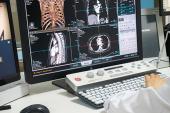
Persistent MR Changes but No Structural Damage
These new data included the subjects of Puntmann et al’s original study, plus additional patients with mild COVID-19 who underwent CMR imaging a median of 109 days after the initial infection. The baseline characteristics of the recovered COVID-19 patients were compared with those of 95 healthy controls.
Among the 346 patients who recovered from COVID-19, 252 (73%) reported cardiac symptoms at baseline, with most having mild (38%) or moderate (33%) symptoms. The symptoms included exertional dyspnea (62%), palpitations (28%), atypical chest pain (27%), and syncope (3%).
Compared with the controls at baseline, COVID-19 patients had higher diastolic blood pressure and myocardial mapping values. They also had more nonischemic myocardial scar by late gadolinium enhancement (LGE), detectable pericardial effusion without hemodynamic relevance, and pericardial enhancement by gadolinium contrast uptake. There was no difference in any of the biomarkers, including C-reactive protein (CRP), high-sensitivity troponin, and N-terminal pro-brain natriuretic peptide (NT-proBNP). They also had no evidence of structural heart disease.
Follow-up CMR was then performed a median of 329 days after the COVID-19 diagnosis, at which time 198 patients (57%) reported cardiac symptoms. Symptoms persisted in 182 individuals from the baseline assessment to follow-up, while 16 previously asymptomatic patients developed new symptoms, 78 asymptomatic patients remained asymptomatic, and 70 patients became asymptomatic during follow-up. Women were more likely than men to experience persistent symptoms (67% vs 46%; P < 0.001).
Exertional dyspnea was the commonly reported symptom, with patients reporting difficulties in getting back to a previous fitness level, challenges climbing stairs or inclines, or limited physical capabilities in professional or everyday life. The more affected patients reported staying home because they feared the onset of general physical weakness, dizziness, or even blackouts.
In terms of the big picture, I think this is actually quite reassuring. Marc Dweck
In the overall COVID-19 cohort, heart rate, systolic blood pressure, and native T1 on CMR tended to decline between baseline and follow-up, but there were also signs that the presence or persistence of symptoms appeared to track with CMR findings. Although native T2 declined and right ventricular ejection fractions increased for the cohort overall, in the patients who remained symptomatic there was a trend toward higher T2 mapping values (reflecting persistent inflammatory edema) than in those who became asymptomatic. In multivariate modeling, female gender and increased native T1 at baseline, reflective of more myocardial fibrosis, were independent predictors of cardiac symptoms at follow-up.
The higher native T2 mapping in those with lingering cardiac symptoms suggest that while inflammation after COVID-19 might be a “pathophysiological commonality” shared by all patients, it may be more pronounced in those with persistent symptoms. To TCTMD, Puntmann said the reason for higher T2 mapping values in those with persistent symptoms is uncertain. One hypothesis is that it might relate to changes in vascular, cellular, or interstitial permeability.
“We believe that what we’re seeing are changes at the small-vessel level, which then creates leaky vessels,” she said. “This may be the source of water that goes into the myocardium.”
Puntmann stressed that this is not myocarditis. As she and her co-authors point out in the paper, “Profound myocardial injury or structural heart disease is not a prerequisite for the presence of symptoms defying the classical definitions of viral myocarditis.”
Limitations to Research
For Dweck, the significance of the higher T2 mapping values in those with persistent symptoms is unclear, particularly since the differences seen were small. “In the absence of any clear difference in the structure or function of the heart, the jury’s out as to what this means,” he said. “So, from my point of view, I do think there are limitations to this study. It’s an interesting paper and a good contribution, but we need to interpret it with caution.
The researchers acknowledge the limitations to their work, notably that selection bias can’t be excluded given that patients referred themselves for screening. As a result, extrapolating these results—the prevalence of symptoms and CMR findings—to the general population of COVID-19 patients is not possible. Additionally, there is significant variability and no standardization when it comes to CMR mapping techniques, making it difficult to extrapolate these results to a real-world setting, said Puntmann.
Puntmann said the follow-up assessments were welcomed by the patients, particularly since many of them didn’t entirely understand what had happened to them following the COVID-19 diagnosis. For the researchers, the findings were equally informative as they confirmed that the cardiac abnormalities seen early were also evident in follow-up.
“We were very early with the first paper, and we wanted to have a little bit more validation of our findings, as well,” said Puntmann. The researchers are continuing to follow these patients and hope to publish 3-year follow-up data when available. “We hope that it’ll provide even more information,” she said.

Michael O’Riordan is the Managing Editor for TCTMD. He completed his undergraduate degrees at Queen’s University in Kingston, ON, and…
Read Full BioSources
Puntmann VO, Martin S, Shchendrygina A, et al. Long-term cardiac pathology in individuals with mild initial COVID-19 illness. Nature Med. 2022;Epub ahead of print.
Disclosures
- Puntmann reports speaking fees from Bayer and Siemens and educational grants from Bayer and NeoSoft.
- Dweck reports no conflicts of interest.
Related News


Comments