Hospital Culture, Physician Preference Mostly Drive IVUS/OCT in PCI
Wide variability was seen, with individual usage among the 334 interventional cardiologists ranging from 0 to 100% of all PCIs.


(UPDATED) Intracoronary imaging to optimize outcomes continues to be used infrequently in the majority of US PCI procedures, but there is a surprising amount of variability at the individual physician and hospital level, according to registry data.
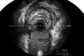
Although RCTs and observational studies have supported the addition of IVUS to optimize PCI, its use is known to be generally low in the US, with one recent study showing just 5.6% of all PCIs in Medicare patients are performed with IVUS guidance. OCT use, too, is rare.
The new study looked at the rate of intracoronary imaging to optimize PCI in a large state registry in Michigan. Lead study author Ryan D. Madder, MD (Frederik Meijer Heart & Vascular Institute, Spectrum Health, Grand Rapids, MI), said while some prior studies had been unable to differentiate between imaging done for optimization and that performed for the purpose of assessing stenosis severity prior to proceeding to PCI, the registry data allow for a clearer picture. The investigators also wanted to see if more-recent studies of imaging had had any impact on IVUS or OCT use.
“We are encouragingly seeing an increase in the overall use of intracoronary imaging to guide PCI,” Madder told TCTMD. “It was 7.2% of all PCI at the beginning of the study and by the last month of the study, it was up to 22.6%.” The difference was statistically significant (P < 0.0001).
But Madder and colleagues also found that among the 48 hospitals included in the analysis, some reported using IVUS or OCT in less than 1% of all PCIs, while others had rates up to 75%.
“When we included the performing hospital and the performing physician as variables in the analysis, the physician performing the PCI, or the hospital performing the PCI, were stronger predictors of whether intracoronary imaging was used compared to every other variable except left main PCI,” Madder added. “That finding represents opportunity for improvement because ideally, we would want clinical factors and evidence-based medicine driving the decisions to perform PCI guided by imaging, not just the hospital or physician that's doing it.”
Commenting on the study for TCTMD, Pinak Shah, MD (Brigham and Women’s Hospital, Boston, MA), said the situation reminds him of the early days of radial artery access.
“Once a couple of physician champions showed that we could do this quickly and patients love it, it just became infectious throughout the lab,” he said. “These findings with regard to intracoronary imaging speak to the staff's familiarity with the technology and to the lab being nimble in getting the device prepped and ready to go and getting rapid image-acquisition interpretation.”
The Influence of Cath Lab Culture
For the study, Madder and colleagues examined data on 48,872 PCIs included in the Blue Cross Blue Shield of Michigan Cardiovascular Consortium (BMC2). The registry includes all patients in the state who undergo PCI (mean age 66 years; 32% female). For the purpose of the study, procedures were included if they were performed between July 2019 and March 2021.
I think one of the things this shows is that we need to work to shift cultures in the cath lab from one where the cath lab accepts the fact that the majority of cases are performed without imaging . . . to one where cath labs become used to performing imaging in the majority of cases. Ryan D. Madder
Intracoronary imaging optimization was used in 8,094 procedures (16.6%). The median hospital frequency was 8.8%, and the median physician frequency was 6.1%. Like the wide variability seen with hospitals, individual usage among the 334 interventional cardiologists ranged from none to 100% of all procedures. On the individual hospital level, use of intracoronary imaging was higher in those with versus without on-site cardiac surgery (17.4% vs 11.5%; P < 0.001).
In a Bayesian analysis, left main PCI was the strongest independent predictor of intracoronary imaging to guide PCI (adjusted OR 4.41; 95% CI 3.82-5.10), followed by proximal left anterior descending artery PCI, in-stent restenosis, and surgical consult prior to PCI. By anatomical location, distal and nondistal left main PCI were both independent predictors. Factors that independently predicted nonuse of intracoronary imaging in PCI were prior CABG, STEMI, left circumflex artery PCI, new-onset angina, NSTE ACS, and increasing age. Further adjustment for patient and procedural characteristics showed that even among physicians with a high rate of use of intracoronary imaging, their individual use rate matched the rate of use of the hospital at which they performed the PCI, such that these physicians had lower rates of use at hospitals with low rates of use. That trend was also seen among physicians with low and intermediate use of intracoronary imaging.
The researchers say the results suggest that cath lab culture has a heavy influence on imaging modality. “I think one of the things this shows is that we need to work to shift cultures in the cath lab from one where the cath lab accepts the fact that the majority of cases are performed without imaging . . . to one where cath labs become used to performing imaging in the majority of cases,” Madder added.
Shah agreed. “If the physician is traveling between hospitals and doing a case at a location where the staff is not up on use of the imaging—they have to go find the machine somewhere, there's a lot of groaning, or whatever—I suspect a busy clinician is probably going to be less likely to use it or really push to try to use it in that situation,” he said.
Use of intracoronary imaging was associated with greater radiation doses, including greater fluoroscopy time, air kerma, and dose area product compared with cases where it wasn’t used. No difference was seen between groups for mortality or other outcomes including stent thrombosis.
One limitation of the study was that the authors were not able to assess the breakdown between IVUS and OCT use, although they say they suspect the majority of the cases involved IVUS. Another limitation is that registry data still leave some uncertainty as to whether the imaging was used to guide stent sizing or to ensure optimal stent expansion.
Madder said the study, like others before it, illustrate just how low the rates of use of IVUS and OCT are in the United States compared with places such as Japan where it is used in 80% or more of PCIs. One thing that might change that, however, is if intracoronary imaging becomes a quality metric, he noted, although more studies are needed to justify that it improves quality and outcomes.
“The data are fairly clear that we do improve long-term PCI outcomes with intravascular imaging, so I think that it should be a quality metric,” Shah said. “In our own lab we do track individual operators use of intracoronary imaging as a metric that displays the data anonymously . . . so that people know exactly where they stand compared to their peers. I think that helps drive self-education and increased usage.”

L.A. McKeown is a Senior Medical Journalist for TCTMD, the Section Editor of CV Team Forum, and Senior Medical…
Read Full BioSources
Madder RD, Seth M, Sukul D, et al. Rates of intracoronary imaging optimization in contemporary percutaneous coronary intervention: a report from the BMC2 registry. Circulation: Cardiovasc Intv. 2022;15:e012182.
Disclosures
- Madder reports receiving research support, speaker honoraria, and consulting fees from Infraredx and speaker honoraria from Abbott Vascular as well as serving on the advisory board of Spectrawave.
- Shah reports receiving research support and consulting fees/speaker honoraria from Edwards and Medtronic.
Related News

Comments