Real-life Patients Do Well With Intravascular Lithotripsy: Spanish Registry
The 30-day registry data bolster findings from pivotal trials of intravascular lithotripsy in calcified coronary lesions.


Intravascular lithotripsy (IVL) is a feasible and safe way to facilitate stenting in severely calcified lesions, even in more-complex cases, 30-day data from the REPLICA-EPIC18 Spanish registry suggest.
Longer follow-up is needed but the early signs are good, said lead author Oriol Rodriguez-Leor, MD, PhD (Hospital Universitari Germans Trias i Pujol, Badalona, and Instituto de Salud Carlos III, Madrid, Spain).
The new results were published online recently in JACC: Cardiovascular Interventions.
“Previous publications with data on IVL came from very short series of patients or [from] selected, low-risk patients” without acute coronary syndromes and lesions that were relatively simple, he told TCTMD. “Our study has the advantage of being prospective. It’s in all-comers, consecutive patients.”
Because “IVL is quite expensive in Spain,” the device was mostly used in the registry after other options had been tried but not worked, Rodriguez-Leor noted. “So it was really a quite complex set of patients and a really complex set of lesions.” With that in mind, the results with IVL in REPLICA-EPIC18 were “amazing,” he said. When asked for their feedback, participating operators reported to investigators that “in more than half of patients, the procedure would have been impossible without IVL.”
The Shockwave Coronary IVL system (Shockwave Medical) has been available in Europe for several years, after the product received CE Mark approval in 2017. In the United States, the Food and Drug Administration approved the Shockwave C2 IVL system in 2021. The device has been greeted by enthusiasm from operators, with some reluctance due to its price tag—mere months later, the Centers for Medicare & Medicaid Services addressed this with a new technology add-on payment that cleared the way for better reimbursement.
Additional data from DISRUPT CAD III, whose 30-day results formed the basis of the FDA’s decision, has since shown sustained benefit up to 1 year after IVL. Promising 1-year results also have been reported by the DISRUPT PAD III trial of peripheral artery disease.
One-year follow-up from REPLICA-EPIC18 is complete, though the data haven’t yet been released, Rodriguez-Leor added. “The results are quite good also.”
Based on what they know now, Rodriguez-Leor said that operators at his center have moved to a lower threshold for when to use IVL. During the registry, it was “only for those cases where there was nothing else we could do,” he added.
REPLICA-EPIC18
REPLICA-EPIC18 prospectively enrolled 426 consecutive patients (n = 456 calcified lesions) treated with IVL across 26 centers, with angiographic analyses and event adjudication done by an independent core lab. The registry had only three exclusion criteria: patient refusal to participate in the study, life expectancy less than 1 year, and hemodynamic instability with Killip class III or IV. Nearly two-thirds of the patients presented with ACS.
Prior to IVL, just 51% of lesions were considered dilatable and numerous tools were used, to varying degrees and in different combinations, for lesion preparation. These included semicompliant, noncompliant, high-pressure, cutting, and scoring balloons as well as rotational atherectomy and, less commonly, orbital or excimer laser atherectomy.
Once the decision was made to use IVL, however, delivery was 99% successful. The primary effectiveness endpoint of procedural success (successful IVL delivery, final diameter stenosis < 20%, and absence of in-hospital MACE) was reached in two-thirds of patients, with no difference between patients with ACS and those with chronic coronary syndromes (CCS; 68% and 65%, respectively; P = 0.47). Angiographic success also was similar in the two groups; notably, there was residual stenosis ≥ 20% in 30.8% of cases.
By 30 days, the MACE rate was 3% overall, 5% in ACS patients, and 1% in CCS patients (P= 0.073).
“It is important to note that the durability of the clinical benefits associated with IVL-optimized stent implantation should be determined in a longer-term clinical follow-up,” the study authors conclude.
Another Piece in the Puzzle for Calcified Lesions
Michael P. Savage, MD, and David L. Fischman, MD (both from Thomas Jefferson University Hospital, Philadelphia, PA), along with Mamas A. Mamas, BMBCh, DPhil (Keele University, Stoke-on-Trent, England), point out in an accompanying editorial that 6% to 20% of patients undergoing PCI have lesions with severe calcification.
The DISRUPT CAD III trial of IVL lacked a comparator arm, and its designs were based on the ORBIT II single-arm, nonrandomized investigational device exemption trial of orbital atherectomy. Thus, the editorialists agree REPLICA-EPIC18 provides valuable insight into IVL’s performance.
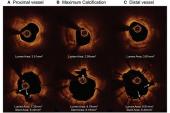
However, they note a few drawbacks to the registry, including the fact that intravascular imaging, which is “particularly important in defining the extent of calcification, in guiding the choice of the calcium modification device, and in assessing vessel size to ensure that an optimally sized IVL balloon is used,” was done before PCI in just 43% of cases.
In addition, the registry had suboptimal angiographic results compared with DISRUPT CAD III, possibly because of the higher-risk cases but also perhaps due to the fact that REPLICA-EPIC18 did not mandate postdilatation, they suggest. “It is likely that many stents were underexpanded, although in the absence of imaging data, it is unclear if this was related to failure to fracture calcium or calcific nodules that might have been better treated with ablative technologies. Because residual stenosis and stent underexpansion are common harbingers of late thrombosis and restenosis, longer-term follow-up of these patients will be important.”
All the more reason, then, to use intravascular imaging both at the outset of PCI and at the end of the procedure, Savage et al assert.
As new tools continue to emerge for addressing coronary calcium, “further research is needed to compare the outcomes of alternative devices, to better harness intravascular imaging to guide device selection, and to explore potential synergistic benefits of combining multiple devices,” they conclude

Caitlin E. Cox is Executive Editor of TCTMD and Associate Director, Editorial Content at the Cardiovascular Research Foundation. She produces the…
Read Full BioSources
Rodriguez-Leor O, Cid-Alvarez AB, Lopez-Benito M, et al. A prospective, multicenter, real-world registry of coronary lithotripsy in calcified coronary arteries: the REPLICA-EPIC18 study. J Am Coll Cardiol Intv. 2024;Epub ahead of print.
Savage MP, Fischman DL, Mamas MA. Between a rock and a hard place: technological progress in treating calcified coronary lesions. J Am Coll Cardiol Intv. 2024;Epub ahead of print.
Disclosures
- The study sponsor, Fundación EPIC, has received an institutional research grant from Shockwave Medical to cover the design and maintenance costs of the electronic case report form. Shockwave Medical has had no influence on the study design or protocol in any respect and was not involved in conducting the research.
- Rodriguez-Leor has received speaker honoraria and consulting fees from Medtronic and World Medica.
- The editorialists report no relevant conflicts of interest.
Related News


Comments